| Alanine, aspartate and glutamate metabolism |    |
| Nitrogen metabolism |    |
| Arginine and proline metabolism |    |
| Glutathione metabolism |    |
| Glycine, serine and threonine metabolism |    |
| One carbon pool by folate |    |
| beta-Alanine metabolism |    |
| Lysine degradation |    |
| Purine metabolism |    |
| Tyrosine metabolism |    |
| Tryptophan metabolism |    |
| Valine, leucine and isoleucine degradation |    |
| Nicotinate and nicotinamide metabolism |    |
| Propanoate metabolism |    |
| Citrullinemia Type I |    |
| Carbamoyl Phosphate Synthetase Deficiency |    |
| Argininosuccinic Aciduria |    |
| Phenylalanine and Tyrosine Metabolism |    |
| Cysteine Metabolism |    |
| Transcription/Translation |    |
| Histidine metabolism |    |
| Amino Sugar Metabolism |    |
| Urea Cycle |    |
| Aspartate Metabolism |    |
| Glutamate Metabolism |    |
| Arachidonic Acid Metabolism |    |
| Piroxicam Action Pathway |    |
| Acetylsalicylic Acid Action Pathway |    |
| Etodolac Action Pathway |    |
| Ketoprofen Action Pathway |    |
| Ibuprofen Action Pathway |    |
| Rofecoxib Action Pathway |    |
| Diclofenac Action Pathway |    |
| Sulindac Action Pathway |    |
| Celecoxib Action Pathway |    |
| Ketorolac Action Pathway |    |
| Suprofen Action Pathway |    |
| Bromfenac Action Pathway |    |
| Indomethacin Action Pathway |    |
| Meloxicam Action Pathway |    |
| Mefenamic Acid Action Pathway |    |
| Oxaprozin Action Pathway |    |
| Nabumetone Action Pathway |    |
| Valdecoxib Action Pathway |    |
| Naproxen Action Pathway |    |
| Glucose-Alanine Cycle |    |
| Malate-Aspartate Shuttle |    |
| 2-Hydroxyglutric Aciduria (D And L Form) |    |
| 2-Methyl-3-Hydroxybutryl CoA Dehydrogenase Deficiency |    |
| 3-Hydroxy-3-Methylglutaryl-CoA Lyase Deficiency |    |
| 3-Methylglutaconic Aciduria Type I |    |
| 3-Methylglutaconic Aciduria Type III |    |
| 3-Methylglutaconic Aciduria Type IV |    |
| 5-Oxoprolinuria |    |
| Adenosine Deaminase Deficiency |    |
| Adenylosuccinate Lyase Deficiency |    |
| AICA-Ribosiduria |    |
| Alkaptonuria |    |
| Beta-Ketothiolase Deficiency |    |
| Canavan Disease |    |
| Dihydropyrimidine Dehydrogenase Deficiency (DHPD) |    |
| Gamma-Glutamyltransferase Deficiency |    |
| Glutaric Aciduria Type I |    |
| Guanidinoacetate Methyltransferase Deficiency (GAMT Deficiency) |    |
| Hawkinsinuria |    |
| Histidinemia |    |
| Hypoacetylaspartia |    |
| Malonic Aciduria |    |
| Maple Syrup Urine Disease |    |
| Methylmalonic Aciduria |    |
| Methylmalonic Aciduria Due to Cobalamin-Related Disorders |    |
| Molybdenum Cofactor Deficiency |    |
| Ornithine Transcarbamylase Deficiency (OTC Deficiency) |    |
| Phenylketonuria |    |
| Prolidase Deficiency (PD) |    |
| Prolinemia Type II |    |
| Purine Nucleoside Phosphorylase Deficiency |    |
| Sialuria or French Type Sialuria |    |
| Tyrosinemia Type I |    |
| Xanthine Dehydrogenase Deficiency (Xanthinuria) |    |
| Non Ketotic Hyperglycinemia |    |
| Propionic Acidemia |    |
| 3-Methylcrotonyl Coa Carboxylase Deficiency Type I |    |
| Isovaleric Aciduria |    |
| Saccharopinuria/Hyperlysinemia II |    |
| Salla Disease/Infantile Sialic Acid Storage Disease |    |
| Dimethylglycine Dehydrogenase Deficiency |    |
| 4-Hydroxybutyric Aciduria/Succinic Semialdehyde Dehydrogenase Deficiency |    |
| Sarcosinemia |    |
| Diflunisal Action Pathway |    |
| Lactic Acidemia |    |
| Glutathione Synthetase Deficiency |    |
| Hyperinsulinism-Hyperammonemia Syndrome |    |
| Pyruvate Carboxylase Deficiency |    |
| GABA-Transaminase Deficiency |    |
| Primary Hyperoxaluria Type I |    |
| Leukotriene C4 Synthesis Deficiency |    |
| Argininemia |    |
| Hyperprolinemia Type II |    |
| Hyperprolinemia Type I |    |
| Arginine: Glycine Amidinotransferase Deficiency (AGAT Deficiency) |    |
| Ornithine Aminotransferase Deficiency (OAT Deficiency) |    |
| Lesch-Nyhan Syndrome (LNS) |    |
| Gout or Kelley-Seegmiller Syndrome |    |
| Tyrosinemia Type 2 (or Richner-Hanhart syndrome) |    |
| Tyrosinemia Type 3 (TYRO3) |    |
| Methylmalonate Semialdehyde Dehydrogenase Deficiency |    |
| Homocarnosinosis |    |
| Tay-Sachs Disease |    |
| Azathioprine Action Pathway |    |
| Mercaptopurine Action Pathway |    |
| Disulfiram Action Pathway |    |
| Thioguanine Action Pathway |    |
| Methotrexate Action Pathway |    |
| Dimethylglycine Dehydrogenase Deficiency |    |
| Hyperglycinemia, non-ketotic |    |
| Ureidopropionase Deficiency |    |
| Carnosinuria, carnosinemia |    |
| Tyrosinemia, transient, of the newborn |    |
| Dopamine beta-hydroxylase deficiency |    |
| Beta-mercaptolactate-cysteine disulfiduria |    |
| 5-oxoprolinase deficiency |    |
| Gamma-glutamyl-transpeptidase deficiency |    |
| Malonyl-coa decarboxylase deficiency |    |
| Creatine deficiency, guanidinoacetate methyltransferase deficiency |    |
| Hyperornithinemia with gyrate atrophy (HOGA) |    |
| Hyperornithinemia-hyperammonemia-homocitrullinuria [HHH-syndrome] |    |
| L-arginine:glycine amidinotransferase deficiency |    |
| Xanthinuria type I |    |
| Xanthinuria type II |    |
| 3-hydroxyisobutyric acid dehydrogenase deficiency |    |
| 3-hydroxyisobutyric aciduria |    |
| Isobutyryl-coa dehydrogenase deficiency |    |
| Isovaleric acidemia |    |
| Hyperlysinemia I, Familial |    |
| Hyperlysinemia II or Saccharopinuria |    |
| Monoamine oxidase-a deficiency (MAO-A) |    |
| G(M2)-Gangliosidosis: Variant B, Tay-sachs disease |    |
| Adenine phosphoribosyltransferase deficiency (APRT) |    |
| Mitochondrial DNA depletion syndrome |    |
| Myoadenylate deaminase deficiency |    |
| Methylenetetrahydrofolate Reductase Deficiency (MTHFRD) |    |
| Succinic semialdehyde dehydrogenase deficiency |    |
| Pyridoxine dependency with seizures |    |
| Warburg Effect |    |
| Antipyrine Action Pathway |    |
| Antrafenine Action Pathway | 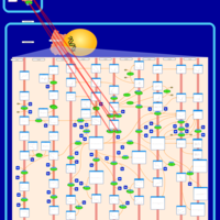   |
| Carprofen Action Pathway |  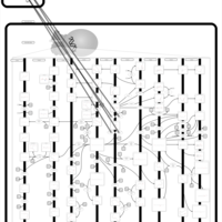  |
| Etoricoxib Action Pathway |    |
| Fenoprofen Action Pathway |    |
| Flurbiprofen Action Pathway |    |
| Magnesium salicylate Action Pathway |    |
| Lumiracoxib Action Pathway |    |
| Lornoxicam Action Pathway |    |
| Phenylbutazone Action Pathway |    |
| Nepafenac Action Pathway |    |
| Trisalicylate-choline Action Pathway |    |
| Tolmetin Action Pathway |    |
| Tiaprofenic Acid Action Pathway |    |
| Tenoxicam Action Pathway |    |
| Salsalate Action Pathway |    |
| Salicylate-sodium Action Pathway |    |
| Salicylic Acid Action Pathway |    |
| Acetaminophen Action Pathway |    |
| 2-aminoadipic 2-oxoadipic aciduria |    |
| 3-Phosphoglycerate dehydrogenase deficiency |    |
| Cystinosis, ocular nonnephropathic |    |
| Folate malabsorption, hereditary |    |
| The oncogenic action of 2-hydroxyglutarate |    |
| Glutaminolysis and Cancer |    |
| The oncogenic action of L-2-hydroxyglutarate in Hydroxygluaricaciduria |    |
| The oncogenic action of D-2-hydroxyglutarate in Hydroxygluaricaciduria |    |
